Contact information
Nuffield Division of Clinical Laboratory Sciences (NDCLS), John Radcliffe Hospital, Oxford
Analysis of Bone Marrow Fibrosis in Blood Cancer
Daniel Royston
MBChB BMSC DPhil FRCPath
Associate Professor of Pathology
- Academic Pathologist
- Consultant Haematopathologist
Quantiative Image analysis of blood cancers using AI / machine learning
Quantiative Image analysis of blood cancers using AI / machine learning
I am an academic Haematopathologist holding a joint University & NHS position. I trained in Medicine & Cellular Pathology before completing my DPhil at the University of Oxford in 2011.
The main focus of my research group is the systematic characterisation of the cellular and stromal interactions within biopsy material that drive the initiation and progression of myeloid blood cancers, with specific focus on myeloproliferative neoplasms (MPN). Our work uses cutting edge techniques in quantitative image analysis, including artificial intelligence (AI) / machine learning approaches. We have successfully demonstrated the utility of these approaches in clinical samples taken from patients, and offered new insights into the fundamental pathological processes driving disease progression and response to therapy. Our work is now employed in the evaluation of several new therapeutics in MPN, and provides clinical trialists with the first robust and objective measures of tissue-based disease modification. Understanding the spatial relationships of key cellular and stromal tissue components and their correlation to genetic and immune / inflammatory factors in myeloid diseases is now the key focus of my research group.
My research involves close multi-disciplinary collaborations between Cellular Pathology, Haematology and Biomedical Engineering (Institute of Biomedical Engineering; Professor Jens Rittscher). Our work is jointly funded by Blood Cancer UK (BCUK) and Cancer Research UK (CRUK).
Publications
Quantitative interpretation of bone marrow biopsies in MPN—What's the point in a molecular age? Ryou H, et al. BJH. 2023. http.org/10.1111/bjh.19154
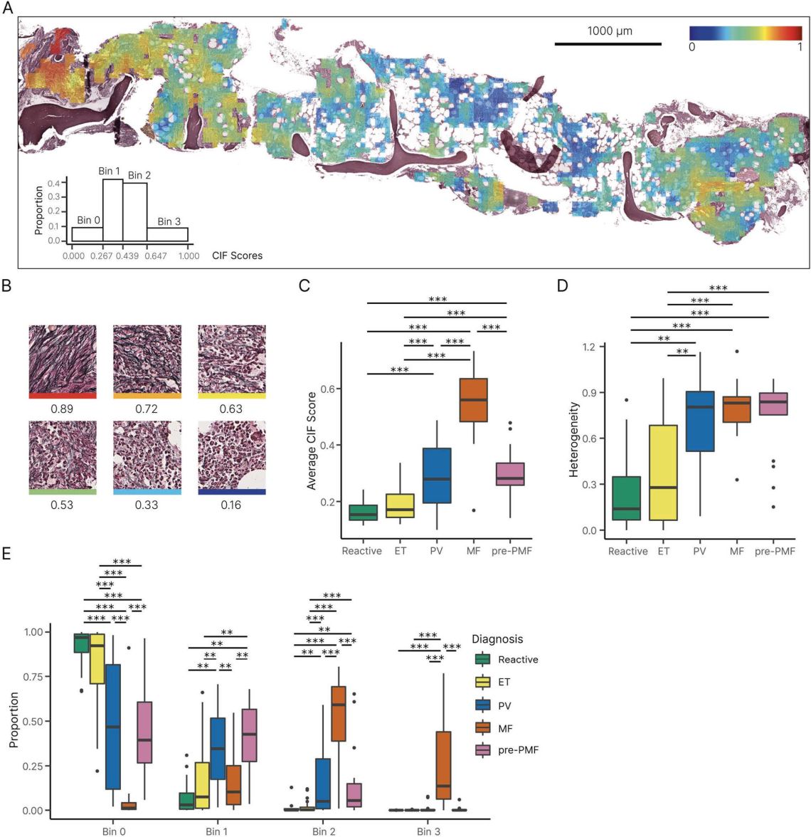
Continuous Indexing of Fibrosis (CIF): improving the assessment and classification of MPN patients. Ryou H, et al. Leukemia. 2022 Dec 5. doi: 10.1038/s41375-022-01773-0.
Application of Single-Cell Approaches to Study Myeloproliferative Neoplasm Biology. Royston D, et al. Hematol Oncol Clin North Am. 2021 Apr;35(2):279-293. doi: 10.1016/j.hoc.2021.01.002.
AI-Based Morphological Fingerprinting of Megakaryocytes: a New Tool for Assessing Disease in MPN Patients. Sirinukunwattana K, et al. Blood Adv. 2020 Jul 28;4(14):3284-3294. doi: 10.1182/bloodadvances.2020002230.
Perspective: Pivotal translational hematology and therapeutic insights in chronic myeloid hematopoietic stem cell malignancies. Mughal TI, et al. Hematol Oncol. 2022 Apr 3. doi: 10.1002/hon.2987. PMID: 35368098.
Multi-Scale Graphical Representation of Cell Environment. Theissen, et al. 2022; 3522-3525.10.1109/EMBC48229.2022.9871710.
Learning Cellular Phenotypes through Supervision. Theissen, et al, 2021. Annu Int Conf IEEE Eng Med Biol Soc. 2021 Nov;2021:3592-3595. doi: 10.1109/EMBC46164.2021.9629898. PMID: 34892015.
In utero origin of myelofibrosis presenting in adult monozygotic twins. Sousos, et al. Nat Med. 2022 Jun;28(6):1207-1211. doi: 10.1038/s41591-022-01793-4.
Single-Cell Analyses Reveal Megakaryocyte-Biased Hematopoiesis in Myelofibrosis and Identify Mutant Clone-Specific Targets. Psaila B, et al. Mol Cell. 2020 May 7;78(3):477-492.e8. doi: 10.1016/j.molcel.2020.04.008.
Collaborators
-
Adam Mead
Professor of Haematology
-
Bethan Psaila
Professor of Haematology
Websites
-
Quantitative Biological Imaging Group / Ludwig Cancer Research
Prof. Jens Rittscher
-
Institute of Biomedical Engineering
Department of Engineering
- Nuffield Division of Clinical Laboratory Sciences (NDCLS)
Recent publications
-
A reconfigurable arbitrary retarder array as complex structured matter.
Journal article
He C. et al, (2025), Nat Commun, 16
-
Delineating Mpl-dependent and -independent phenotypes of Jak2 V617F- positive MPNs in vivo.
Journal article
Papadopoulos N. et al, (2025), Blood
-
Reticulin-Free Quantitation of Bone Marrow Fibrosis in MPNs: Utility and Applications.
Journal article
Ryou H. et al, (2025), EJHaem, 6
-
Entering the era of spatial transcriptomics: opportunities and challenges for pathology
Journal article
Sozanska AM. et al, (2025), Diagnostic Histopathology
-
The spleen
Chapter
Hildyard C. and Royston D., (2025), Hoffbrand S Postgraduate Haematology Eighth Edition, 71 - 81
-
Spatial transcriptomic approaches for characterising the bone marrow landscape: pitfalls and potential.
Journal article
Cooper RA. et al, (2024), Leukemia
-
A proinflammatory stem cell niche drives myelofibrosis through a targetable galectin-1 axis.
Journal article
Li R. et al, (2024), Sci Transl Med, 16
-
Clinical utility of investigations in triple-negative thrombocytosis: A real-world, multicentre evaluation of UK practice.
Journal article
Godfrey AL. et al, (2024), Br J Haematol
-
Quantitative analysis of bone marrow fibrosis highlights heterogeneity in myelofibrosis and augments histological assessment: An Insight from a phase II clinical study of zinpentraxin alfa
Journal article
Ryou H. et al, (2024), HemaSphere, 8
-
Nasopharyngeal myeloid sarcoma as a manifestation of acute monomyelocytic leukaemia.
Journal article
Pervaiz A. et al, (2023), BMJ case reports, 16


